weird
A Canine, Drilled Through, With a Lens in It
Yes, and it restores sight. A canine is removed, drilled through, fitted with a lens, grown under the skin for months, and then implanted into the eye.
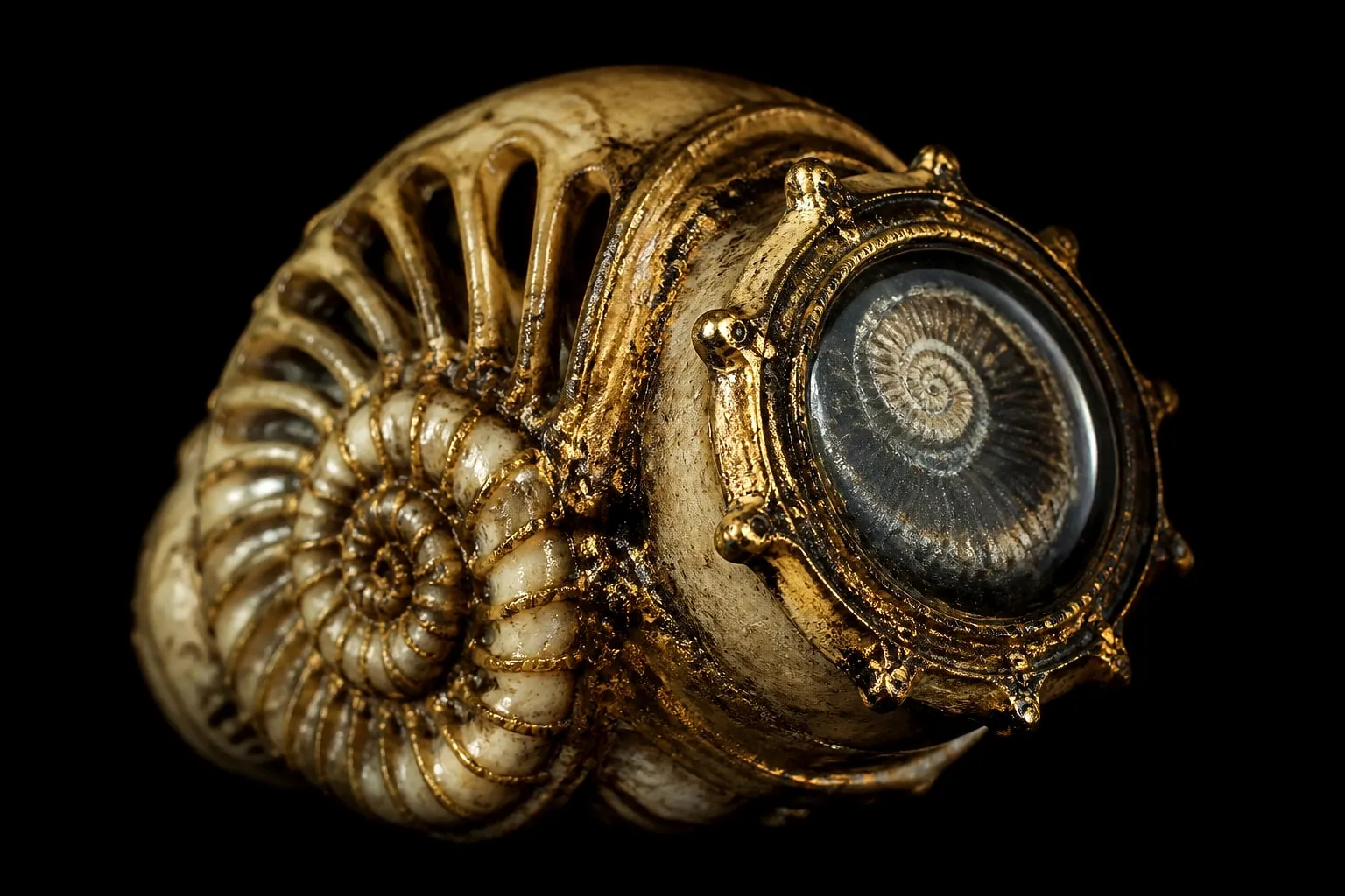
This sounds like an internet fabrication and it is a real operation with sixty years of published outcomes.
It’s called osteo-odonto-keratoprosthesis — bone-tooth-cornea-replacement — and for a small group of people it is the difference between blindness and reading a book.
The problem it solves
The cornea is the clear window at the front of the eye. If it becomes opaque, light cannot get in, and the person is blind even though the retina and optic nerve work perfectly.
Usually the answer is a corneal transplant. But some eyes cannot accept one — after severe chemical burns, after Stevens-Johnson syndrome, in advanced ocular pemphigoid — because the surface of the eye has lost the cells and the tear film a graft needs to survive. A transplant into that environment fails, repeatedly.
The obvious alternative is an artificial window: a plastic cylinder set into the cornea. And that runs into the second problem, which is the interesting one.
The eye extrudes it. Living tissue does not bond to plastic. Over months the body works the implant loose at the edges, and it falls out, taking the eye’s integrity with it.
So the requirement is a frame that living tissue will accept, holding a clear cylinder in the middle.
Which is where the tooth comes in
Benedetto Strampelli, working in Rome in the 1960s, solved it with a piece of the patient.
A canine tooth is chosen — it has the longest single root and the thickest surrounding bone. The tooth is removed together with a block of the jawbone around it and a strip of the ligament that joins them.
That block is then sliced into a flat lamina: a thin plate of dentin with bone on either side.
A hole is drilled through the middle, and a small optical cylinder of PMMA — clear acrylic — is cemented into the hole.
The result is a lens held in a frame made of the patient’s own tooth and jaw.
And then it is grown under the skin
This is the step that makes the operation genuinely strange.
The lamina is implanted into a pocket under the skin of the cheek, or in the eyelid, or in the abdominal wall, and left there for two to four months.
The purpose is to let soft tissue grow into and around the bone, so that when it reaches the eye it arrives already integrated — with its own blood supply and a covering of living tissue rather than as a loose object.
Meanwhile the eye is prepared. The damaged surface is removed, and a graft of the patient’s own cheek lining — buccal mucosa — is laid over the front of the eye. That mucosa is tough, it is used to a wet environment, and it will accept the implant where the original ocular surface would not.
Months later the lamina is retrieved from the cheek and implanted into the prepared eye, with the optical cylinder passing through the mucosa so light can reach the retina.
The result looks nothing like an eye
The patient ends up with a small dark cylinder protruding through pink tissue where a cornea used to be. There is no visible iris, no white, no recognizable eye.
And they can see. Reported outcomes in properly selected patients are strikingly good — many achieve vision good enough to read, and long-term series find the majority of laminae still in place two decades later.
For someone blind for years with no alternative, the cosmetic result is not the part they are weighing.
Why the tooth specifically
Three properties, and no synthetic material has all three.
It is autologous. Taken from the patient, so there is no immune rejection to manage and no donor to wait for.
Dentin is dimensionally stable. It doesn’t resorb the way pure bone grafts often do, and it holds a cemented cylinder without loosening.
And the bone edge integrates. Soft tissue grows onto it and seals around it, which is exactly the thing plastic cannot do and the reason plastic implants extrude.
The tooth is not a poetic choice. It is the only structure in the body that is both hard enough to machine and biologically welcome in the eye.
The version without a tooth
For patients who have no suitable canine — or none at all — the same principle is used with a tibial lamina, a plate cut from the shin bone. It works, and outcomes are generally reported as slightly less durable than the tooth version, which is a strong argument for how good dentin is at the job.
What it says about teeth
We treat teeth as parts of a chewing mechanism, and the moralized version of dental care makes them a report card on somebody’s discipline.
They are neither. They are the hardest, most stable mineralized structure the human body builds — hard enough to grind rock for eighty years, stable enough to be sliced into a plate, drilled, and used as a permanent frame inside an eyeball.
A tooth removed for a cavity and thrown away is the same material as the one restoring somebody’s sight in Rome.
The short version
Osteo-odonto-keratoprosthesis, developed in Italy in the 1960s, still performed today for corneal blindness that no transplant can fix.
A canine and its surrounding jawbone are removed, sliced flat, drilled through, fitted with a clear acrylic cylinder, grown under the skin of the cheek for months, then implanted into an eye resurfaced with cheek lining.
It looks alarming. Most of them are still working twenty years later.
Questions people actually ask
Is tooth-in-eye surgery real?
Yes. It's called osteo-odonto-keratoprosthesis, developed by Benedetto Strampelli in Italy in the 1960s, and it is still performed in a handful of specialist centers for people who cannot receive a conventional corneal transplant.
Why use a tooth?
Because the eye rejects most foreign material, and a plastic lens placed directly in the cornea works loose. A slice of the patient's own tooth and jawbone is not rejected, and living bone bonds to tissue in a way plastic does not.
Which tooth is used?
Usually a canine, because it has the longest single root and the thickest bone around it. The tooth and the surrounding jawbone are removed as one block, sliced, and drilled through the middle for the optical cylinder.
Does it work?
For the right patients, remarkably well. Long-term studies report the majority of implants still in place after twenty years, in people who were completely blind beforehand and had no other option.